Cancer Staging: A Tale of Bias
March 9, 2018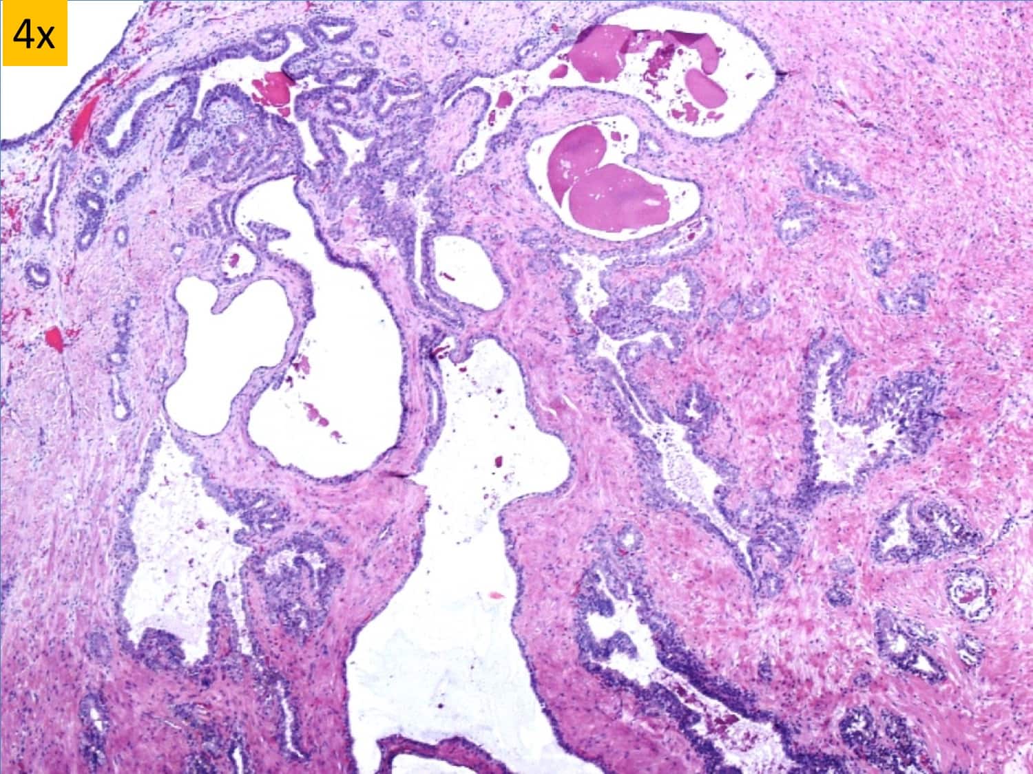
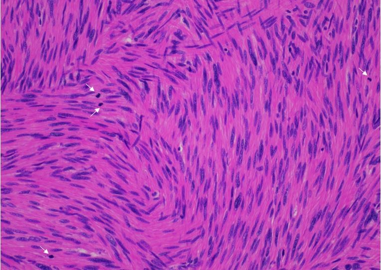
48-year-old man with retrovesical cystic mass
March 19, 2018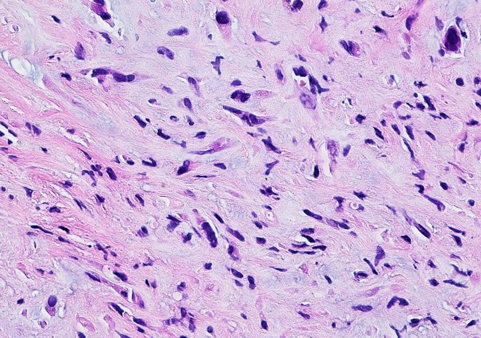

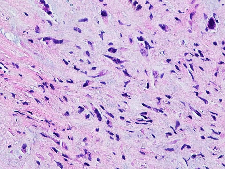

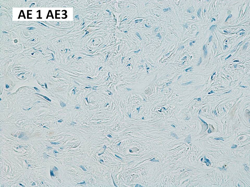

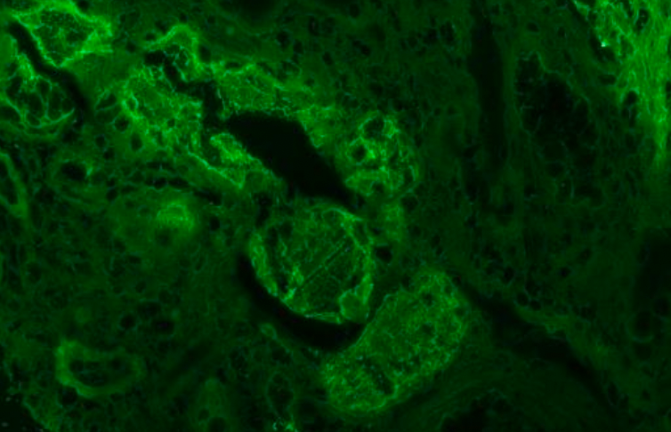
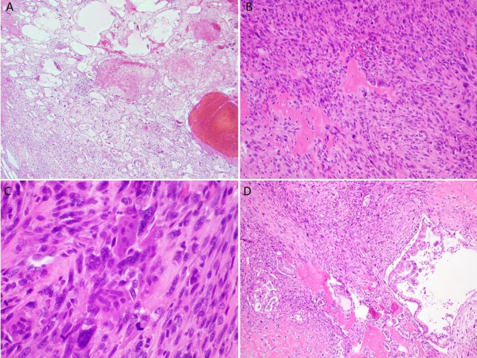
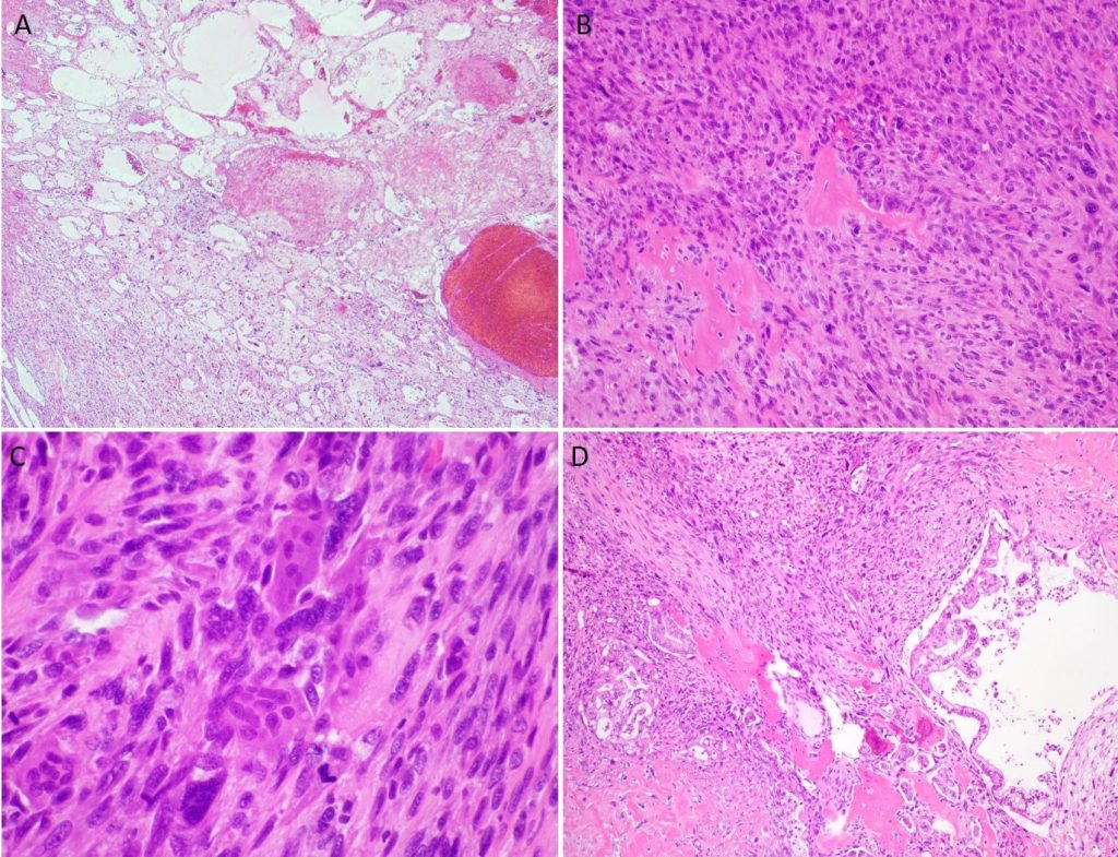
A 31 year old woman presented to an urgent care clinic for cough and nasal congestion which would not resolve with OTC treatment of what appeared to be sinusitis. Chest xray at the urgent care clinic revealed innumerable small nodules. Further imaging revealed lesions 1.5 – 1.8 cm in the posterior and lateral right lobes of her liver. A CT directed biopsy of the liver was performed, and slides of the biopsy are shown. There are epithelioid tumor cells with focal intracytoplasmic vacuoles embedded in a fibromyxoid matrix. The epithelioid cells are rounded with eosinophilic cytoplasm, and mild to moderately atypical nuclei. Immunostains show tumor cells are positive for CD31, CD34, Factor VIII and Fli-1, but negative for pacytokeratin AE1/AE3, CK7 and TTF-1. The Ki-67 labeling index is moderately elevated ( 5-10%). Reticulin and trichrome stains show fibrotic stroma in the tumor. The findings are consistent with Hepatic Epithelioid Hemangioendothelioma.