69-year-old woman with pneumatosis of colon

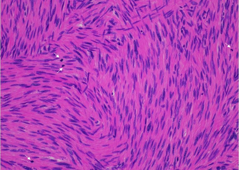
Focus on fibrotic lesions with multinucleated giant cells
March 30, 2018
NYPS President’s Symposium
March 31, 2018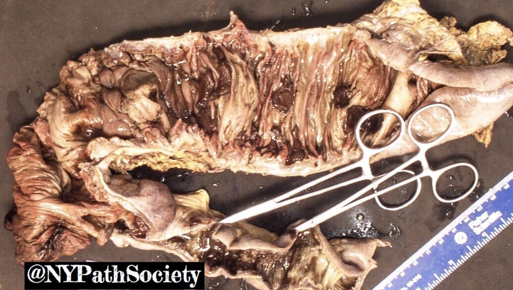
A 69 year old woman on chemotherapy for a neuroendocrine tumor presented to the emergency room with hyponatremia, hyperkalemia and acute kidney injury. She was diagnosed with tumor lysis syndrome, chemotherapy was discontinued, and she received medical therapy to control her electrolyte abnormalities. A CT showing pneumatosis of small bowel and colon prompted urgent exploratory laparotomy with right hemicolectomy for ischemia. In the gross image, the specimen appears dusky gray with brown congested areas. Low and intermediate power photomicrograph of the mucosa show necrotic villi and diffuse inflammation. Of note, purple crystalline material is found diffusely in the necrotic mucosa and luminal debris, these crystals show a fish-scale pattern when examined on light microscopy.
What is your diagnosis?
The diagnosis is Kayexalate (Polystyrene sulfonate) induced colitis. This patient received Kayexalate, which is a common therapy for hyperkalemia. The medication binds intraluminal potassium to prevent further potassium absorption. The medication is known to crystallize in the gastrointestinal tract in patients with kidney injury, causing acute bowel ischemia and necrosis. On detection of these crystals it is important to emergently notify the clinical team that the Kayexalate must be discontinued.
A similar effect is associated with Sevelamer, a resin that is used to treat hyperphosphatemia. However, the crystals associated with Sevelamer are yellow/pink, while Kayexalate crystals are purple. Both Sevelamer and Kayexalate crystals may have fish scale like impressions on light microscopy.
Case submitted by Dr Yonah Ziemba of Zucker School of Medicine at Hofstra/ Northwell Health
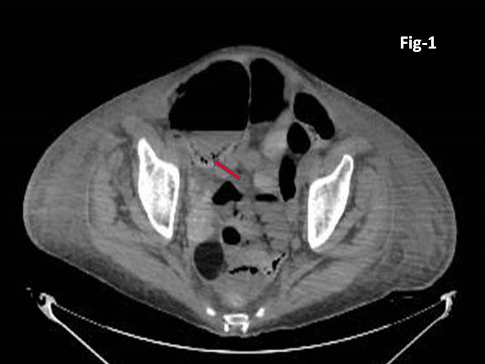
Fig -1 CT scan showing punctate air around the bowel (arrow heads) as pneumatosis coli. In addition there are early air fluid levels suggesting bowel stasis.
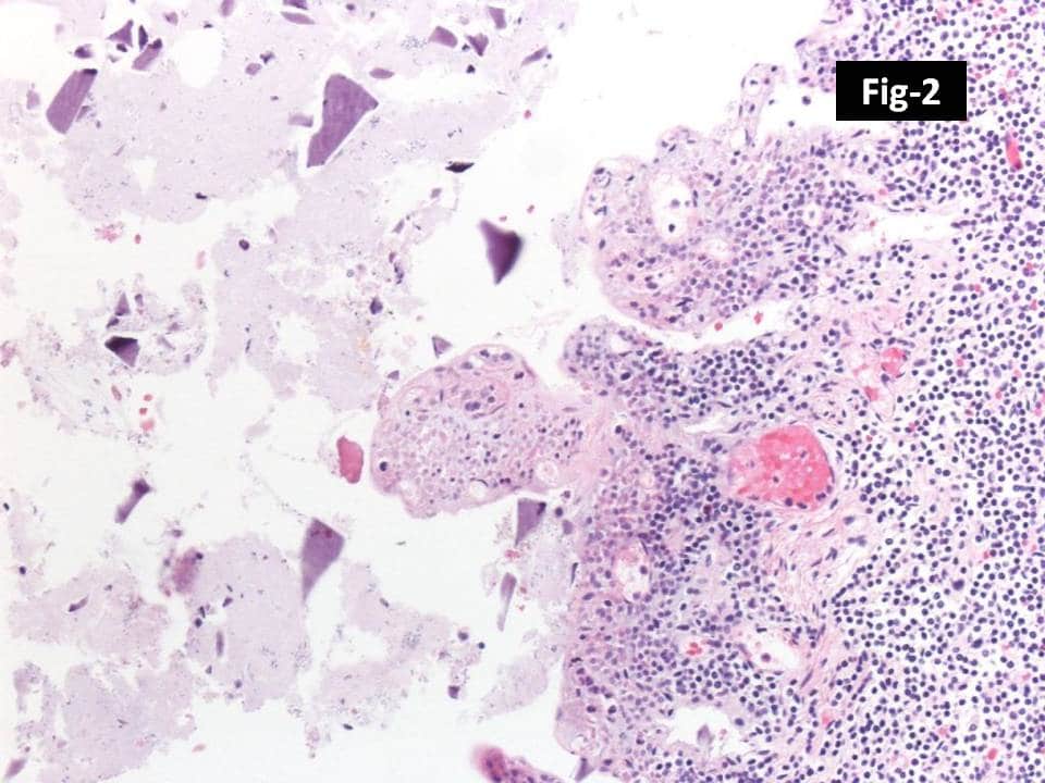
Fig – 2 Necrotic small bowel with impression of necrotic villi and luminal debris with crystalline material (x40).
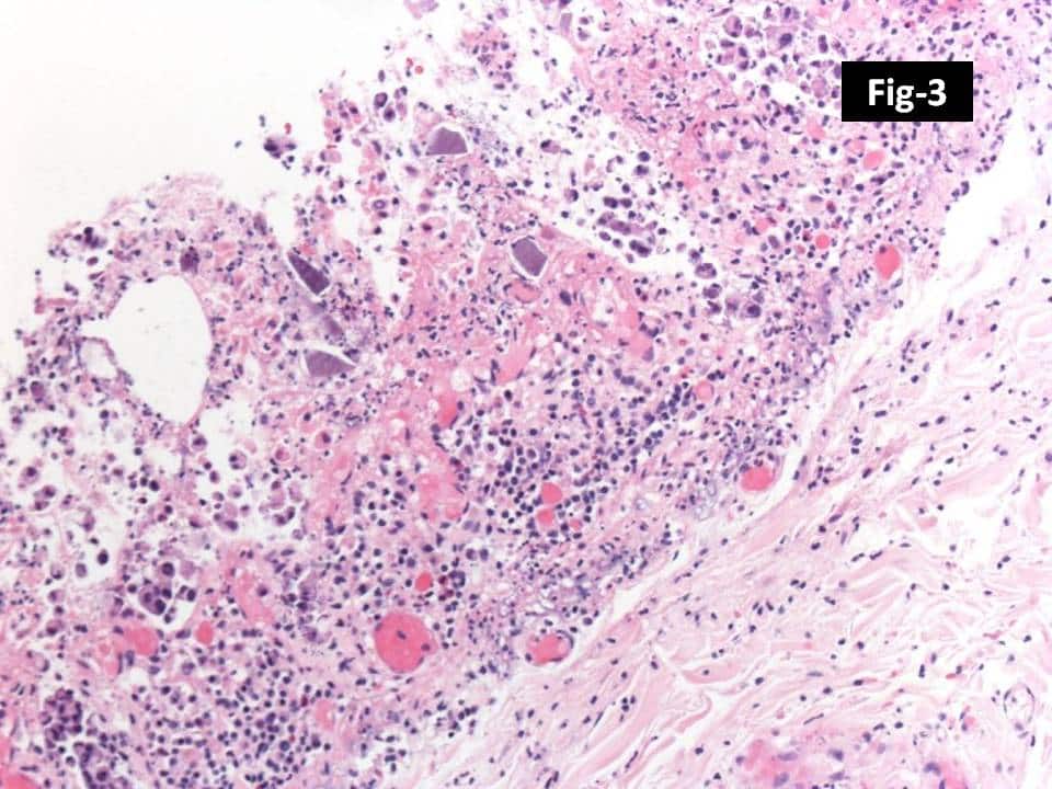
Fig – 3 Extensive areas of mucosal necrosis with crystalline material within the necrotic mucosa (x100).
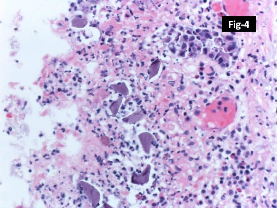
Fig – 4 Higher magnification of the purple-violet crystalline material (x200).
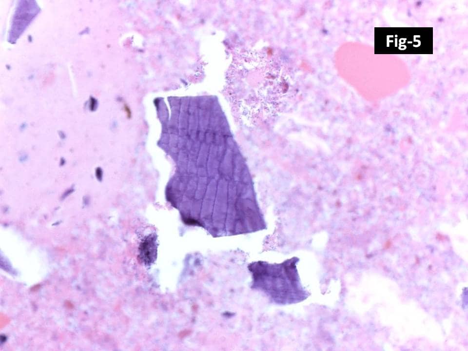
Fig – 5 Higher magnification of purple-violet crystalline material demonstrating “fish-scale” like configuration in background of necrotic debris (x400).