48-year-old man with retrovesical cystic mass

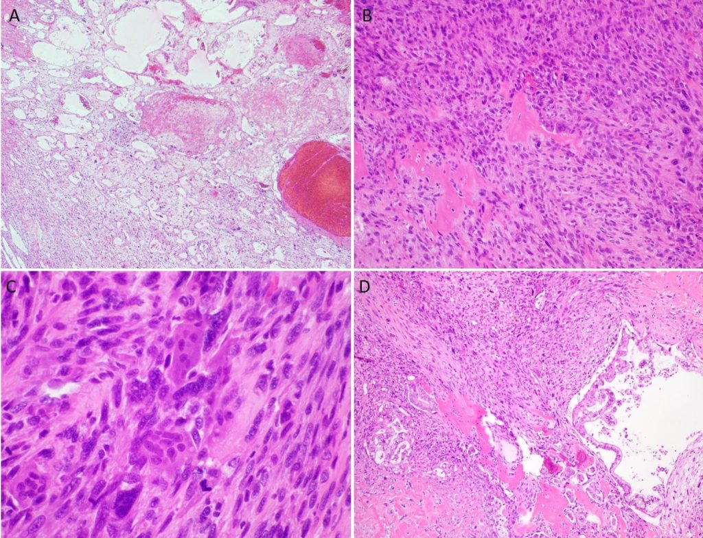
31-year-old woman with liver mass
March 10, 2018Bacteriology Board Review
March 29, 2018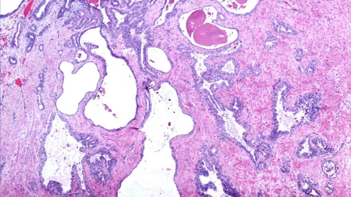

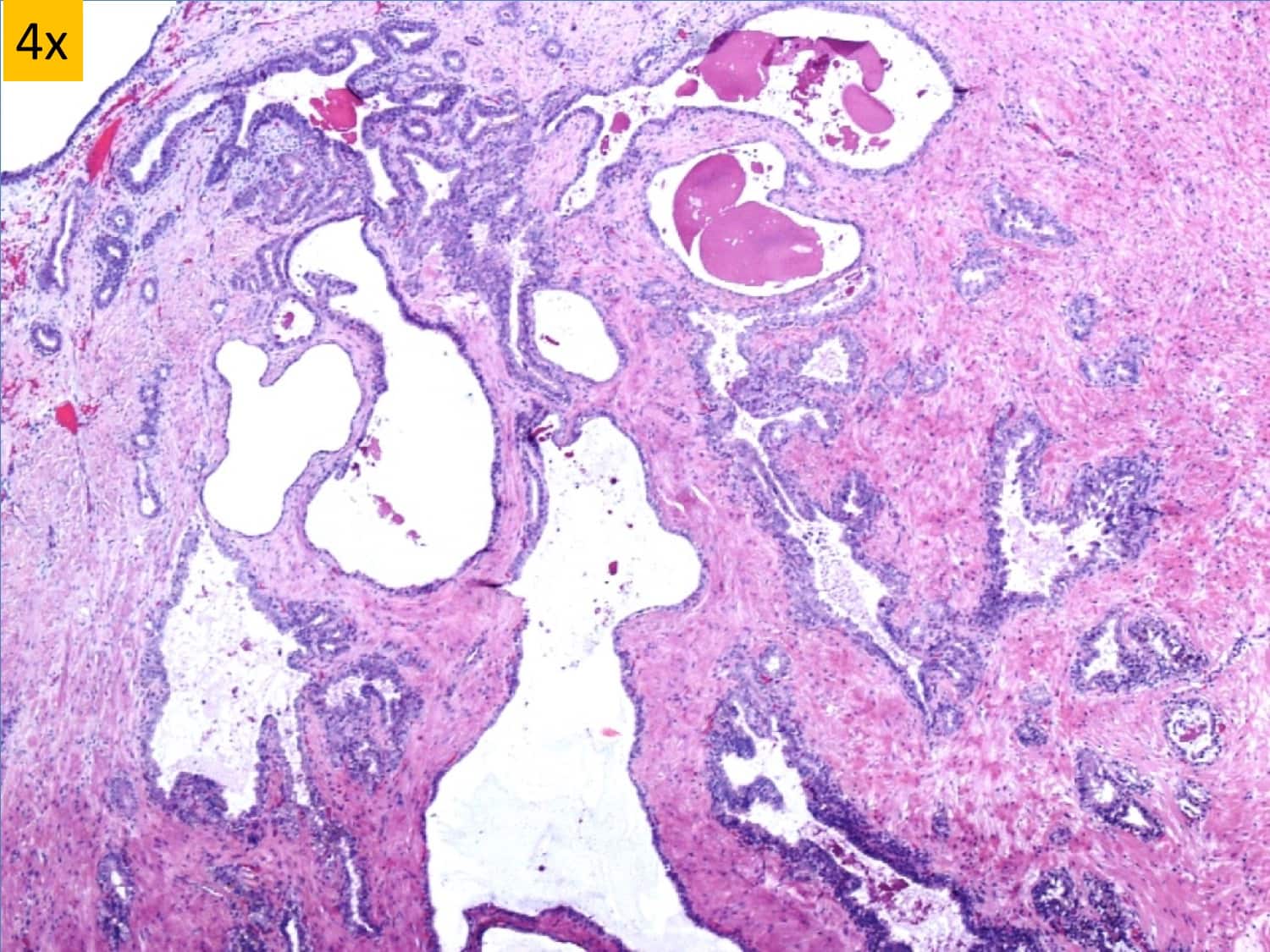
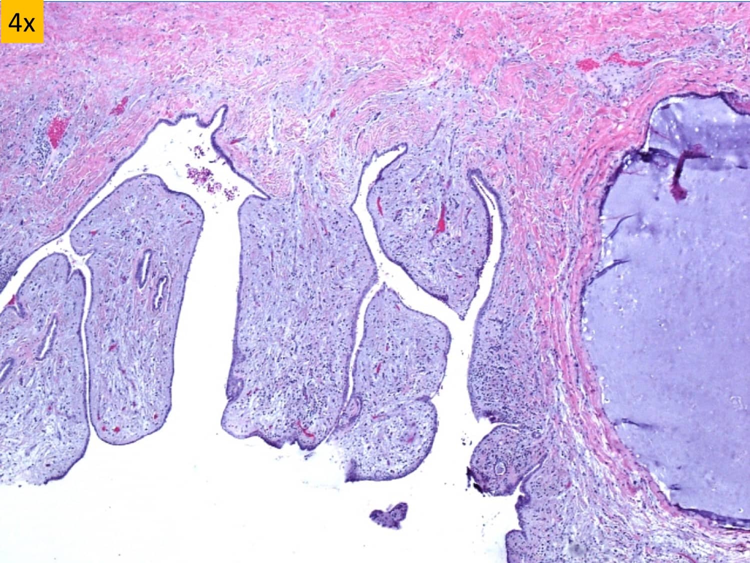
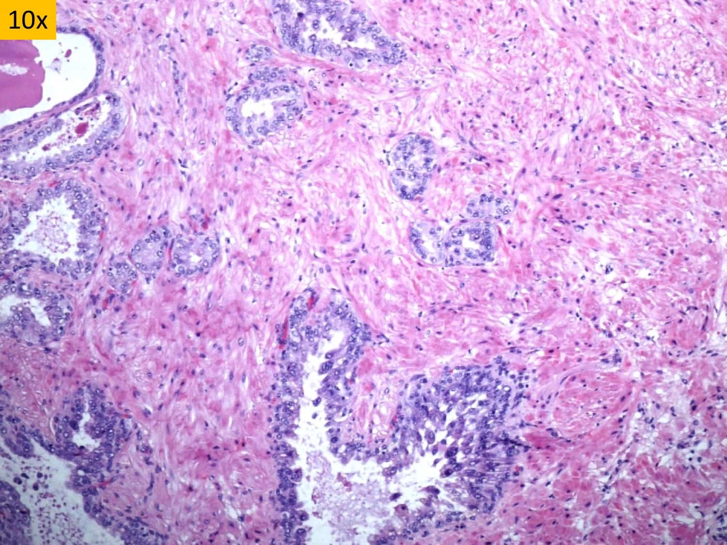
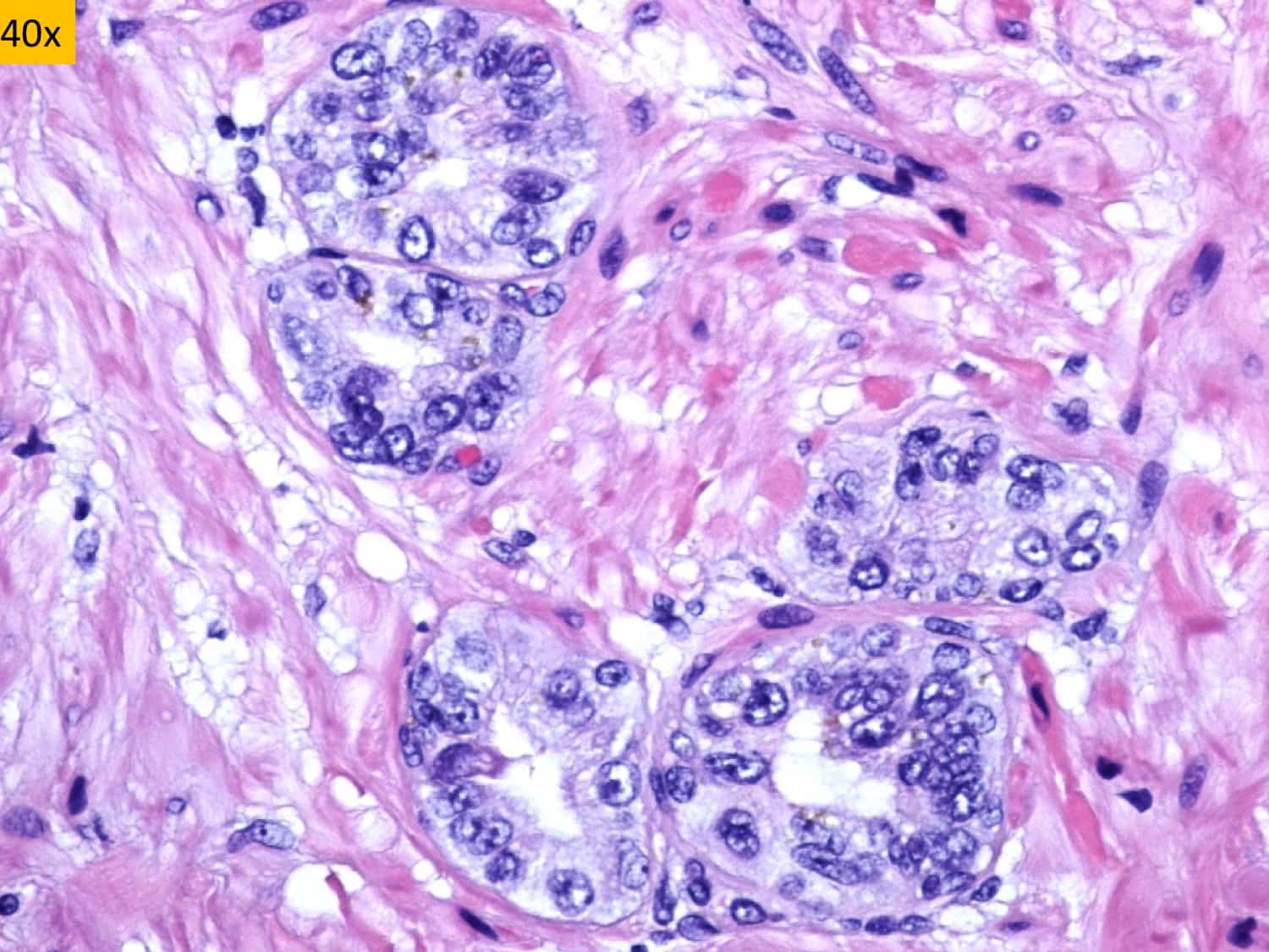
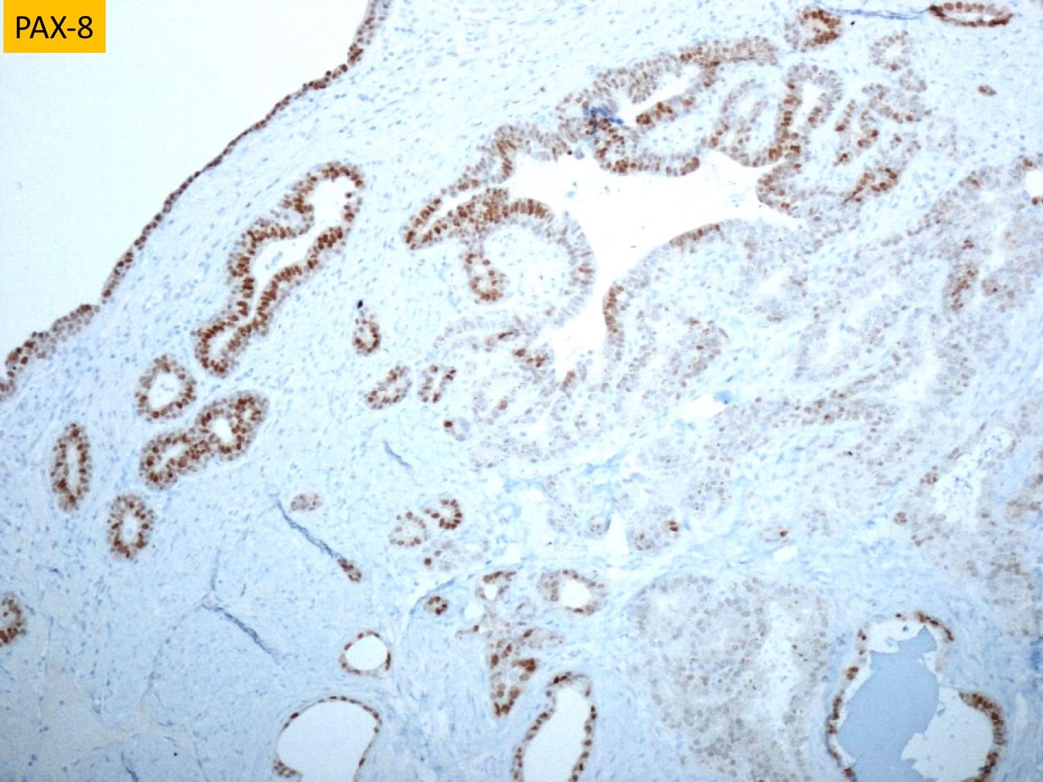
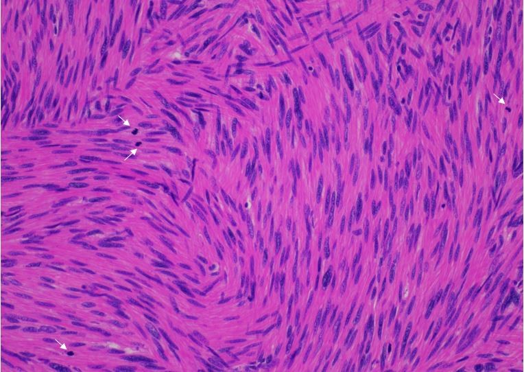
A 48 year-old male with a history of congenital left renal agenesis and hydronephrotic right kidney was found to have 11 cm asymptomatic retrovesical cystic mass on routine follow-up CT scan of abdomen and pelvis. The cyst was excised. Grossly, it was a 10 cm thick-walled cyst with a tan-brown to pink-purple shaggy outer surface. The inner surface was tan-brown with smooth, bumpy and vaguely papillary areas with several small cysts containing transparent mucoid material. Histologically, the cyst consisted of hypocellular fibromuscular wall lined by epithelium of variable thickness. The epithelial lining ranged from single layer of flat/cuboidal cells with squamoid morphology to columnar mucin containing cells to multilayer of cells with clear to basophilic cytoplasm. Luminal broad-based papillations and small intramural cysts containing amorphous proteinaceous to mucoid material were present. Significant stromal or epithelial atypia, mitoses, nuclear pleomorphism, or necrosis were not identified (images shown). Immunohistochemically, the epithelial lining was positive for PAX-8 (image shown) and negative for PSA, PSAP and WT-1. The final diagnosis is most consistent with cystadenoma of seminal vesicle.
Case submitted by Dr. Neha Gupta, Donald and Barbara Zucker School of Medicine at Hofstra/Northwell.