64-year-old woman presented with uterovaginal prolapse

The Continuing Dilemma of Ductal Carcinoma in situ
January 26, 2018NYPS Residents Forum
February 16, 2018
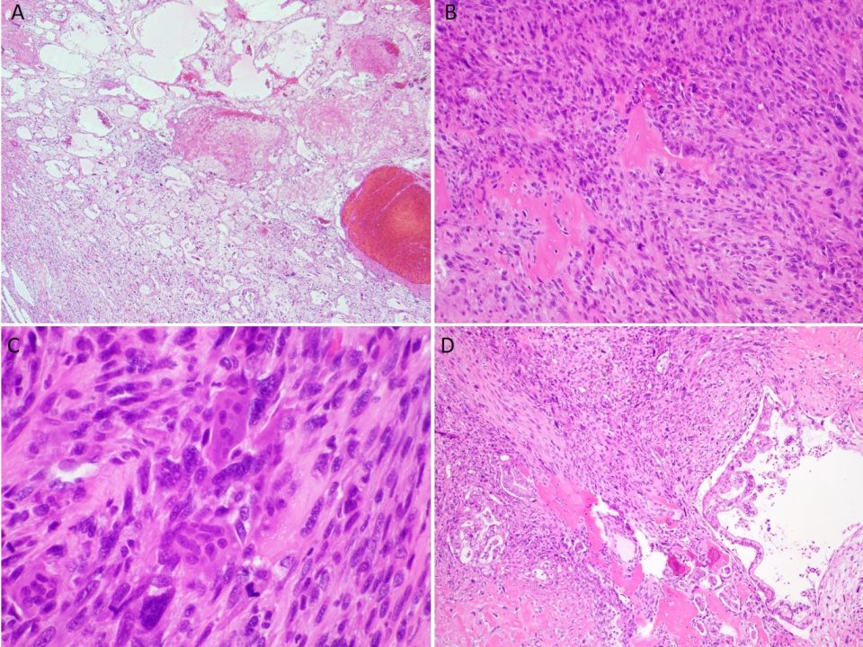
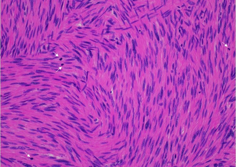
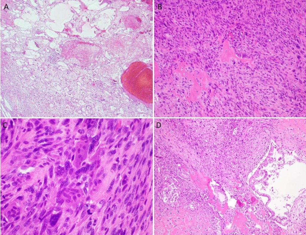
Section of endomyometrium, in a 64 year old woman who underwent total hysterectomy for uterovaginal prolapse.
The endomyometrium show regular, round and focally crowded glands, haphazardly infiltrating the myometrium with no inflammatory or desmoplastic response. The glands contain eosinophilic secretions and simulate mesonephric remnants or mesonephric hyperplasia. The immunohistochemical markers for mesonephric differentiation ( GATA3, calretinin and CD10) are negative. Additionally mCEA is also negative. The glands are positive for PAX8 and ER. This pattern is most consistent with endometrioid adenocarcinoma with an adenoma malignum pattern of myometrial invasion.
Case submitted by Dr. Sonali Lanjewar, Albert Einstein Medical Center
References:
- Longacre TA, Hendrickson MR. Diffusely infiltrative endometrial adenocarcinoma: an adenoma malignum pattern of myoinvasion. Am J Surg Pathol. 1999; 23(1): 69-78.
- Tambouret R, Clement PB, Young RH. Endometrial endometrioid adenocarcinoma with a deceptive pattern of spread to the uterine cervix: a manifestation of stage IIb endometrial carcinoma liable to be misinterpreted as an independent carcinoma or a benign lesion. Am J Surg Pathol. 2003; 27(8): 1080-8.